
![]()
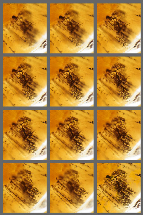
Here is a single nerve cell from the cerebellum of the brain seen using 4 different methods of 3D imaging:
1) A stack of images of a single nerve cell is seen at different focus levels The images here are of the same nerve cell, photographed at 11 different focus levels in the microscope. You will notice that each photograph has some parts that are in focus and others that are out of focus. The 12th image at the end was reconstructed by stacking the 11 previous images providing a resultant image that has all of the in-focus data.
|
 |
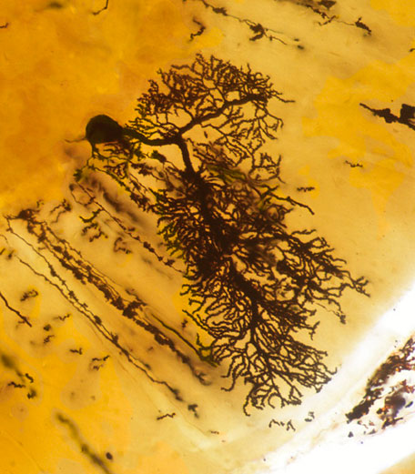
2) A deep-focus image was reconstructed from the stack of 2D optical slices A deep-focus image of the nerve cell was reconstructed from the stack of optical slices above |
 |
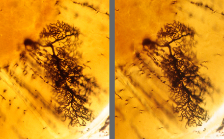
3) A real-time stereo image of the nerve cell at one focus level The two images here are Left and Right eye images for 3D viewing by using cross-eye viewing. |
 |
| 4) A fully focused stereo 3D image of the nerve cell | 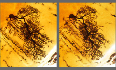 |
5) real-time motion parallax image of nerve cell This Animated GIF clip shows how our patented motion parallax method creates three dimensionality on a two dimensional screen. (Please be patient as this animation may take a few moments to load fully). |
 |
© 2002-2018 sandgrains.com All Rights Reserved